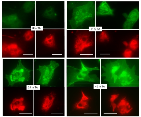
Fig. 3.
Parietal cells expressing ezrin-T567A (TA)-cyan fluorescent protein (CFP) have a similar time course of appearance and distribution of the ezrin-TA mutant as for the ezrin-WT. As in Fig. 1, cell cultures were infected with rAd-ezrin-TA-CFP construct and prepared for immunocytochemistry at indicated sequential times. Expression of ezrin-TA-CFP (green) and H-K-ATPase (red) is shown. Bar markers = 20 μm.

