
FIG. 6.
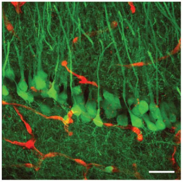
Maximal two-dimensional projection of a three-dimensional reconstruction of deconvolved and merged z-stacks of native red and green fluorescence from 200-μm thick tissue sections using confocal laser-scanning microscopy. The CA1 pyramidal cell layer in the hippocampus from an adult IRG/Nestin-Cre double transgenic mouse is shown. Recombined pyramidal cells are green and nonrecombined blood vessels are red. Scale bar: 20 μm.