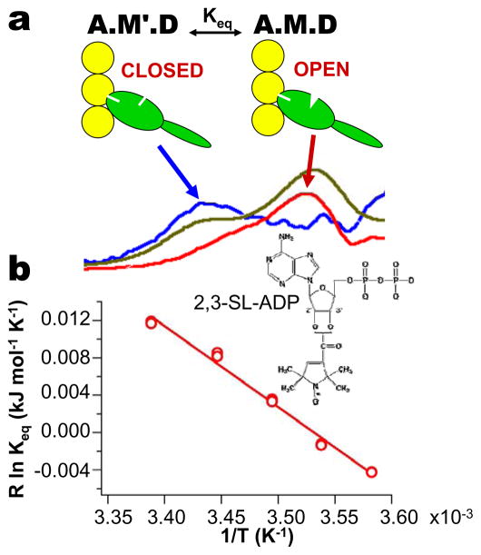
Figure 7.
Structure and thermodynamics of the nucleotide pocket deduced from EPR (48). (a) Low-field portion of EPR spectrum of SL-ADP bound to myosin in rabbit psoas muscle (orange), deconvoluted into its two components (blue and red). (b) van’t Hoff plot of Keq for opening of the pocket, determined from EPR.

