Figure 6.
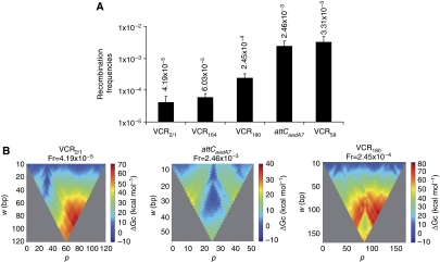
In vivo extrusion of the attC sites secondary structures from double-stranded DNA. (A) Recombination frequencies of different attC sites after transformation of attC-containing plasmids in non-replicative permissive recipient cells (see Materials and methods: recombination assay with a non-replicative substrate). Error bars show s.d. (B) Energy landscape of cruciform formation. The frequency of recombination (Fr) for each attC site is indicated. Each colour dot represents the free energy of cruciform formation (ΔGc in kcal/mol) for a portion of the attC site of length w (bp) at the position p (from favourable in blue to unfavourable in red). The tip of the triangle is the free energy of the whole attC site. As attC sites are symmetrical, favourable energies that are not along the centre of the triangle represent favourable non-recombinogenic structures. attCaadA7 folds in a favourable recombinogenic site (blue colour from the middle of the base of the triangle). VCR2/1 folds in a favourable non-recombinogenic structure (blue colour shifted from the middle). VCR180 presents neither favourable recombinogenic nor favourable non-recombinogenic structures.

