Figure 1.
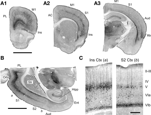
EGFP expression in the forebrain of the 5-HT2AR-EGFP mouse. (A,B) Low magnification images illustrating the distribution of EGFP expressing cells in coronal and horizontal sections. Calibration bar = 2 mm. AC, anterior cingulate cortex; Aud, auditory cortex; Ent, Entorhinal cortex; Hipp, hippocampus; Ins, insular cortex; M1, primary motor cortex; Orb, orbital cortex; PL, prelimbic cortex; Rh, rhinal cortex; S1, primary somatosensory cortex; S2, secondary somatosensory cortex, Str, striatum. Low magnification images of parasagital brain sections for this mouse are available at the GENSAT web site. (C) Higher magnification images depicting the EGFP immunoreactivity in the insular [a in (B)] and secondary somatosensory cortex [b in (B)]. Calibration bar = 250 μm. In this figure EGFP was immunolocalized using a rabbit anti-GFP antibody and detected using immunoperoxidase histochemistry.

