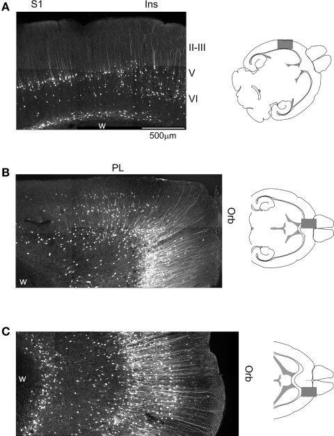
Figure 2.
Cellular expression of EGFP in the somatosensory, prefrontal and orbital cortices of 5-HT2AR-EGFP mice. (A) Confocal image illustrating the distribution of EGFP immunoreactive cells in the primary sensory and adjacent insular cortex. Calibration bar = 500 μm. (B) Distribution of EGFP in the medial prefrontal and medial orbital cortices. (C) Distribution of EGFP in the lateral orbital cortex. Ins, insular cortex; Orb, orbital cortex; PL, prelimbic cortex; S1, primary somatosensory cortex. EGFP was immunolocalized in horizontal sections using a rabbit anti-GFP antibody and detected using an Alexa 488 conjugated secondary goat anti-rabbit antibody. Images depict flattened confocal stacks containing 15–20 optical sections spanning 20–30 μm along the z axis.

