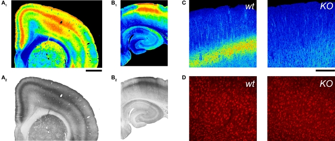
Figure 4.
5-HT2A receptor detection in the cerebral cortex using an anti-5HT2A receptor antibody. (A,B) 5-HT2A receptor immunoreactivity in the anterior cerebral cortex (A) and hippocampus (B) in a wild type mouse. 5-HT2A receptor immunodetection for these images used the ImmunoStar antibody at a 1:100 dilution and HRP/DAB detection (A2,B2). (A1) and (B1) depict pseudocoloring of images in (A2) and (B2).Calibration bar: 1 mm. (C) 5-HT2A receptor immunoreactivity in the cerebral cortex of wild type and 5-HT2A receptor knockout (KO) mice. Notice the disappearance of the layer V band in the KO mouse. 5-HT2A receptor immunodetection for these images used the ImmunoStar antibody at a 1:500 dilution and an Alexa 488 conjugated secondary antibody. Images are pseudocolored for emphasis and depict comparable flattened confocal stacks through motor-frontal cortex. (D) Immunoreactive staining obtained with the Ab51 antibody. Images correspond to single confocal sections through layers V and VI of the M1 region in a wild type and a 5-HT2A receptor knockout mice. (C,D) Calibration bar: 200 μm.

