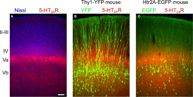
Figure 6.
Laminar distribution of 5-HT2A receptor immunoreactivity in motor-frontal cortex. (A) Laminar distribution of 5-HT2A receptors (red) in mouse cortex counterstained using a fluorescent Nissl dye (blue, Neurotrace, Invitrogen). (B) 5-HT2A receptors (red) distribution in the motor (agranular) cortex of the Thy1-YFP mouse. In this mouse YFP (green) is expressed in the large pyramidal cells of layer Vb. Notice the double banded distribution of YFP expressing cells that characterizes agranular cortex in this mouse (Shepherd, 2009). (C) 5-HT2A receptor distribution in the cortex of the 5-HT2AR-EGFP mouse. Calibration bar: 100 μm.

