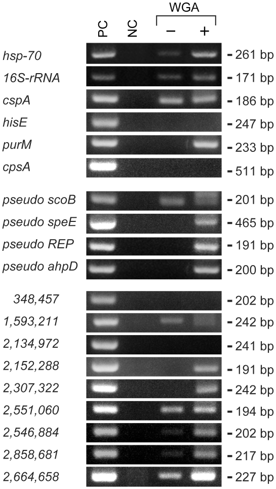
Figure 5. Comparison of conventional PCR and WGA-PCR.
PCR was performed using a DNA sample derived from the present study (sample No. 1 shown in Table 1 and Fig. 3). The DNA before and after WGA (“−” and “+”, respectively) was amplified using specific nested primers and direct primers targeted for the genes, pseudogenes and non-coding regions of M. leprae genome as listed in Table 2. Names of the genes and pseudogenes are indicated and the coordinate positions of non-coding regions in the genome (http://genolist.pasteur.fr/Leproma/) are indicated numerically. PC: positive control DNA from Thai 53 strain of M. leprae; and NC: negative control using DNase/RNase-free water instead of a DNA sample for PCR reaction.

