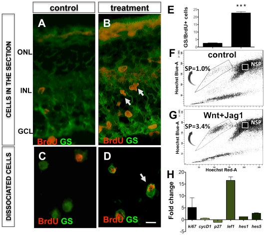
Figure 11. Notch and Wnt signaling-mediated activation of Müller cells in S334ter rats in vivo.
S334ter rats received intravitreal injections of Jag1+Wnt2b at PN10 and PN11, followed by immunofluorescence, Hoechst dye efflux and Q-PCR analyses on PN 13. Immunofluorescence analysis of retinal sections (A, B, arrows) and cell dissociates revealed the presence of BrdU+ cells co-expressing GS (C, D), whose proportion was significantly higher than in controls (E). Hoechst dye efflux assay revealed a higher proportion of SP cells (3.4%) in Jag1+Wnt2b treated retina, compared to that in controls (1%) (F, G). Q-PCR analysis of gene expression revealed increase in levels of transcripts corresponding to Ki67, cyclinD1, Hes1, Hes5 and decrease in p27kip1 transcript levels in Jag1+Wnt2b treated retina, compared to controls (H). ONL = outer nuclear layer; INL = inner nuclear layer; GCL = ganglion cell layer. Scale = 5 µM. *** = p<0.0001.

