Figure 1.
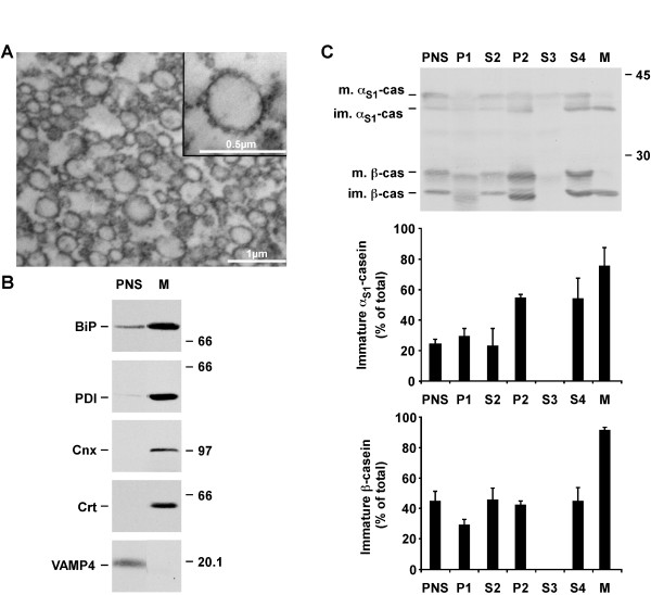
The rough ER microsomal fraction from a lactating rat is almost pure. The rough ER microsomal fraction (M) was prepared from a post-nuclear supernatant (PNS) of mammary tissue from lactating animals by differential centrifugation followed by sucrose density gradient. A. An aliquot of the ER microsomal fraction was fixed and processed for electron microscopy. Insert shows a microsome associated with numerous ribosomes. B. Aliquots of PNS and of the ER microsomal fraction were analyzed by SDS-PAGE followed by immunoblotting for the indicated protein markers. Representative ECL signals from two independent experiments are shown. C. Aliquots of the different supernatants (PNS, S) and pellets (P, M) collected during the purification (see Methods for detailed identification of the fractions) were analyzed by SDS-PAGE followed by immunoblotting using an antibody against mouse milk proteins. The amount of immature forms of αS1- and β-caseins was quantified by densitometry and expressed as percent of the total quantity of the individual casein (mature + immature). The mean ± s.d. of three independent experiments is shown. Relative molecular masses (kDa) are indicated on the right of the immunoblots. αS1-cas: αS1-casein; β-cas: β-casein; BiP: immunoglobulin heavy chain binding protein; Cnx: calnexin; Crt: calreticulin; im.: immature; m.: mature; VAMP4: vesicle-associated membrane protein 4.

