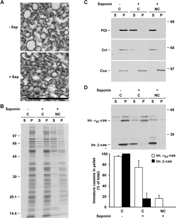
Figure 4.
The majority of αS1-casein, but not of β-casein, remains in the ER after saponin permeabilisation of microsomal membranes in conservative conditions. A. Electron micrographs of microsomes incubated in conservative conditions in the absence (- Sap) or the presence (+ Sap) of saponin and centrifuged. The membrane pellet was fixed and processed for electron microscopy. B-D. Aliquots of the microsomes were diluted and incubated in conservative (C) buffer in the absence of saponin (-), or in conservative or non-conservative (NC) buffers in the presence of saponin (+). After centrifugation, supernatants (S) and pellets (P) were analyzed by SDS-PAGE. B. Coomassie Blue staining. C. Immunoblotting using antibodies against the indicated ER-resident proteins. D. Immunoblotting with an antibody against mouse milk proteins. Immature αS1- and β-casein were quantified by densitometry and the proportion of the immature form in the pellet was expressed as percent of the total quantity of the casein (supernatant + pellet). The mean ± s.d. of three independent experiments is shown. Relative molecular masses (kDa) are indicated. Scale bar: 1 μm. Cnx: calnexin; Crt: calreticulin; im. αS1-cas: immature αS1-casein; im. β-cas: immature β-casein.

