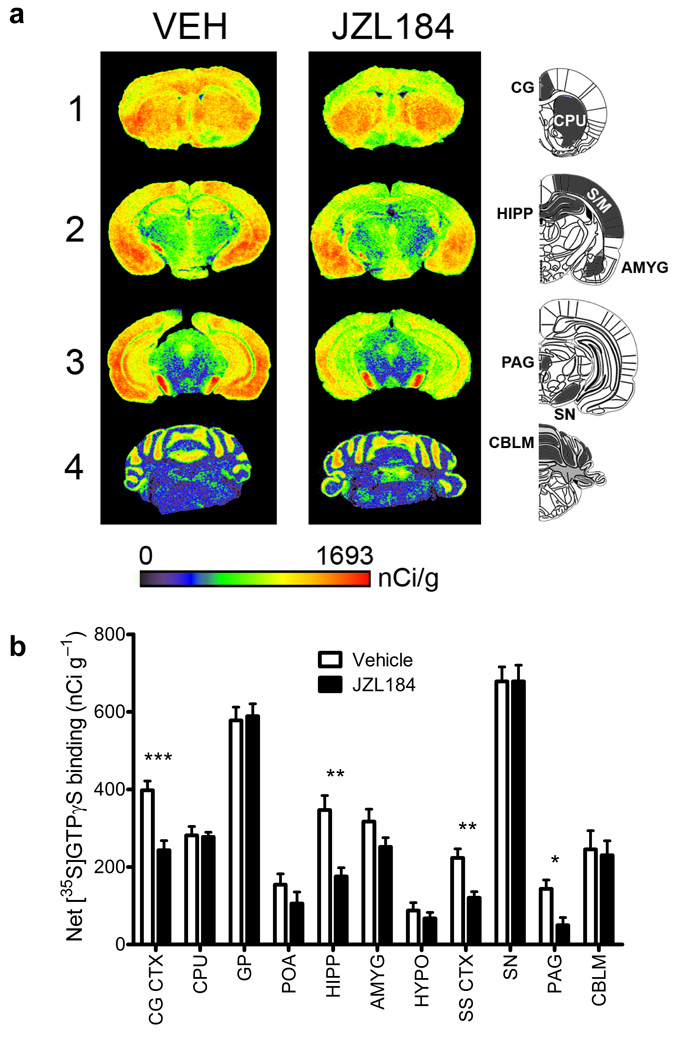
Figure 5. Regional changes in cannabinoid agonist-stimulated [35S]GTPγS binding following chronic disruption of MAGL.
(a) Representative autoradiograms showing CP55,940-stimulated [35S] GTPγS binding in coronal brain sections following either chronic vehicle (left column) or JZL184 (right column) treatment. Pseudocolor images indicate levels of receptor-mediated G-protein activity and highlight significant decreases in CB1 receptor activation in the cingulate cortex (CG, row 1), hippocampus (HIPP, row 2) and periaqueductal gray (PAG, row 3), while no differences are apparent in the caudate putamen (CPU, row 1) or cerebellum (CBLM, row 4). (b) Densitometric analysis of CP55,940-stimulated [35S]GTPγS binding in selected regions, including: cingulate cortex (CG CTX), caudate putamen (CPU), globus pallidus (GP), preoptic area of the hypothalamus (POA), hippocampus (HIPP), amygdala (AMYG), hypothalamus (HYPO), somatosensory cortex (SS CTX), substantia nigra (SN), periaqueductal gray (PAG), & cerebellum (CBLM). Data are presented as means ± s.e.m. n = 8 brains per group, run in triplicate slices for each targeted region. *p < 0.05, **p < 0.01, ***p < 0.001 versus vehicle treatment for specific region (Student’s t-test).

