Figure 7.
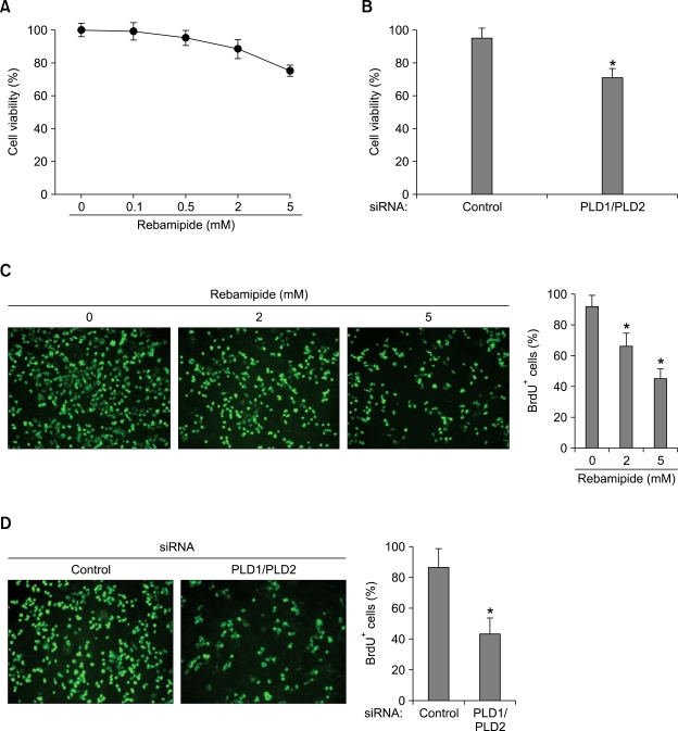
Rebamipide-induced suppression of PLD inhibits the proliferation of gastric cancer cells. (A) MKN-1 cells were treated with the indicated concentrations of rebamipide for 24 h. The cell viability was then measured using the MTT assay. (B) The cells were transfected with both siRNAs for PLD1 and PLD2 for 48 h. The cell viability was measured using the same methods. Each value represents the mean ± S.D. of three independent experiments. *P < 0.05 versus control siRNA. (C) After MKN-1 cells were treated with the indicated concentrations of rebamipide for 24 h, proliferation of the cells was analyzed by BrdU incorporation (green). Scale bar, 100 µm. Quantification of the percentage of BrdU-positive (BrdU+) cells in total living cells of control. *P < 0.05 versus control siRNA. (D) After MKN-1 cells were co-transfected with both siRNAs for PLD1 and PLD2, proliferation of the cells was analyzed by BrdU incorporation (green). Scale bar, 100 µm. Quantification of the percentage of BrdU-positive (BrdU+) cells in total living cells of control-siRNA. *P < 0.05 versus control-siRNA.

