Figure 1.
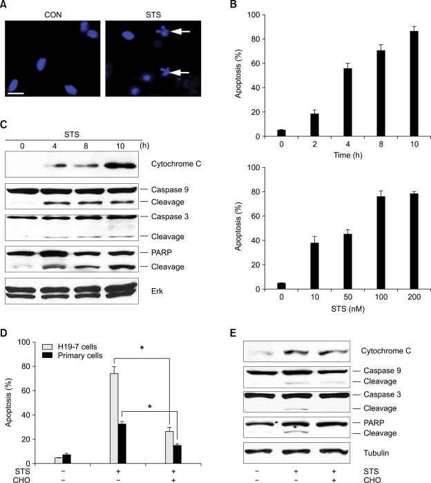
Staurosporine induces neuronal apoptosis. (A) Cultured hippocampal neurons were exposed to 100 nM of STS. After 8 h, apoptotic cell death was determined after nuclei staining with DAPI. All the quantitative analysis of apoptotic nuclei in this report are given as the mean ± standard error (s.e.) from three separate experiments, *P < 0.05 (Student's t-test), and all the pictures in this report were taken using a fluorescent microscope (×100). White arrows indicate apoptotic and fragmented nuclei. Scale bar = 5 µm. (B) H19-7 cells were treated with 100 nM STS from 2 to 10 h (upper) or with STS from 10 nM to 200 nM for 8 hours (lower). Cells were collected at the indicated times and stained with DAPI. (C) H19-7 cells were exposed to 100 nM STS and collected at the indicated times. 30 µg of cell lysate was immunoprobed with indicated antibodies. (D) H19-7 cells and cultured hippocampal cells were untreated or treated with caspase 3 inhibitor CHO for 30 min before exposure to 100 nM STS for 8 hours. (E) H19-7 cells were treated as described in (D) and Tubulin was shown as a loading control.

