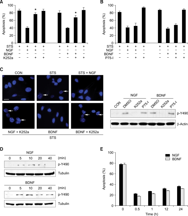
Figure 3.
NGF and BDNF protect neuronal apoptosis through Trk signaling. (A and B) H19-7 cells were pretreated with or without K252a or p75NTR inhibitor for 30 min and incubated with or without 100 ng/ml NGF or BDNF for additional an hour. The cells were then exposed to 100 nM STS in for 8 h. *P < 0.05. (C) (left) Images of Trk inhibitor K252a-blocked NGF and BDNF prevented nuclear condensation. H19-7 cells were treated as indicated, and nuclei were stained with DAPI. White arrows inidicate apoptotic and fragmented nuclei. Scale bar = 5 µm. (right) K252a inhibits phosphorylation of Tyr490. H19-7 cells were treated as indicated and 30 µg of cell lysate was immunoprobed with anti-phospho-Tyr490 antibody. (D) Activation of Trk receptors in H19-7 cells by NGF and BDNF. H19-7 cells were treated with NGF or BDNF at the indicated times. Cell lysates then were probed with anti-phospho-Tyr490 antibody. (E) H19-7 cells were pretreated with 100 ng/ml NGF or BDNF on the indicated times before exposing to STS. Condensed nuclei were counted under the fluorescent microscope and presented under bar graph.

