Figure 1.
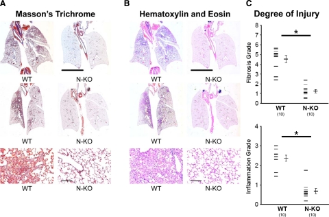
Evaluation of bleomycin-induced lung fibrosis. A and B: Bleomycin-induced lung injury in wild-type (WT) and N-KO mice 14 days after drug administration. Whole lung sections were stained with Masson’s trichrome (A) or H&E (B). These representative sections show focal inflammation and fibrosis in lungs from wild-type mice. In contrast, lungs from N-KO mice show a near-normal appearance. The line represents 5 mm. The lower panels are pictures of lesions taken at a higher magnification (the line is 20 μm). C: Lung histology was graded by a pathologist who was blinded to mouse genotype. Fibrosis was graded using a scale described by Ashcroft et al,24 varying from 0 (normal lung) to 8 (total fibrosis). Inflammation was graded using the 0 (normal lung) to 4 (maximum inflammation) scale described by Sur et al.25 The inactivation of the N-terminal catalytic site of ACE significantly reduced the lung injury scores for both fibrosis and inflammation (n = 10 per group; *P < 0.001).

