Figure 1.
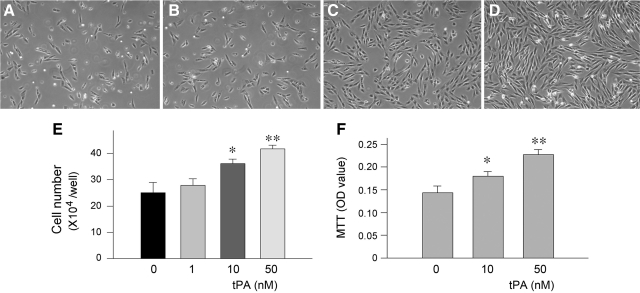
tPA promotes renal interstitial fibroblast proliferation. A–D: NRK-49F cells were incubated with different concentrations of tPA for 48 hours. Representative micrographs show the phase-contrast images of NRK-49F cells after treatment without (A) or with 1 nmol/L (B), 10 nmol/L (C), and 50 nmol/L tPA (D), respectively. E: Cell numbers after treatment for 48 hours with different concentrations of tPA as indicated. F: Cell growth assessed by a colorimetric MTT assay. *P < 0.05, **P < 0.01 versus controls (n = 3).

