
Figure 8.
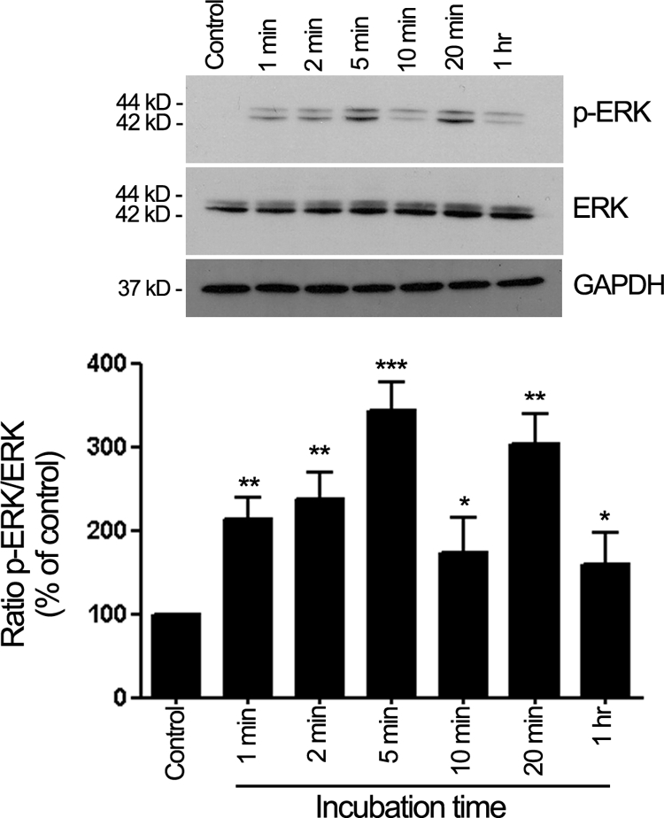
HQ activates ERK1/2 phosphorylation. Time-dependent ERK1/2 phosphorylation assessed by Western blot on cell lysates from ARPE-19 cells treated with 100 μmol/L HQ for various periods of time. Top: Western blot from a representative experiment. Number on the left represents protein molecular weight in kilodaltons. Bottom: Average densitometry results of three independent experiments run in duplicate. Phospho-ERK (p-ERK) protein expression was normalized to total ERK. GAPDH was used as loading control for total ERK. Data are expressed as percentage of control and are means ± SE. *P < 0.05, **P < 0.01, and ***P > 0.001 versus control.