Figure 7.
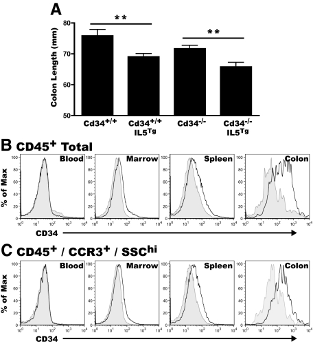
IL5-induced eosinophilia leads to colon shortening, with CD34 expression on colon infiltrating eosinophils. Colon tissues from unchallenged, non-DSS-treated, IL5Tg and Cd34−/−IL5Tg at steady state were excised, measured. and processed for flow cytometry. Blood, bone marrow and spleen tissues were also processed for flow cytometry as described in Materials & Methods. A: Baseline colon length of Cd34+/+ and Cd34−/− animals with or without the IL5 transgene. Histogram profiles of CD34 expression on total CD45+ cells (B) and CD45+/CCR3+SSChi cells (C) in peripheral blood, marrow, spleen, and colon isolates. For all histograms, black lines represent staining on cells from Cd34+/+IL5Tg tissues and gray-filled histograms represent background staining of cells from Cd34−/−IL5Tg tissues. (Histograms are representative of three separate experiments; n = 9 to 12 for colon length; **P < 0.01; Error bars = SEM).

