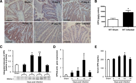
Figure 1.
OPN expression is increased in response to C. rodentium infection. A: Colonic immunohistochemistry showed that OPN was present at low levels in uninfected wild-type (WT) mice (top left panel), mainly located in the cytoplasm of surface epithelial cells. A time-dependent increase in OPN expression was observed throughout the crypts of C. rodentium-infected wild-type mice as the infection progressed to 10 days, followed by a reduction in OPN by PI day 15. Scale bar: 100 μm. B: Enzyme-linked immunosorbent assay of wild-type mouse colons demonstrated a threefold increase in OPN levels with infection (N = 4 to 8; t-test: *P < 0.005). C: Western blotting of colonic homogenates of wild-type mice showed a similar increase in OPN protein level, normalized to β-actin, on PI day 6 (N = 4 to 5; analysis of variance: *P < 0.01) and on PI day 10 (t-test: **P < 0.05). D: Colonic mRNA levels of OPN, relative to β-actin, showed increased OPN expression in infected colons on PI day 10 by qPCR (N = 5 to 9; analysis of variance: *P < 0.01). E: Serum OPN levels, as measured by enzyme-linked immunosorbent assay, were increased on PI day 10 and normalized by PI day 15 (N = 5 to 9; analysis of variance: *P < 0.05).

