Figure 3.
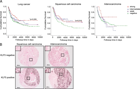
KLF5 expression significantly correlates with better disease-specific survival of human NSCLC patients. KLF5 immunohistochemical analysis was performed on biopsies of 609 human NSCLC tissues on array slides. For disease-specific survival analyses, a total of 258 cases were compared by Mantel-Cox log rank analysis for KLF5 expression levels that had interpretable KLF5 data, follow-up information >30 days, and known cause of death. A: Kaplan-Meier disease-specific survival curves of lung cancer patients of all subtypes (left), squamous cell carcinoma (middle), and adenocarcinoma (right). P values are indicated where statistical significance was found. B: Representative images of KLF5-negative and KLF5-positive immunohistochemical staining of squamous cell carcinomas and adenocarcinomas from the NSCLC tissue array. KLF5 positive cells are dark brown in contrast to KLF5 negative cells and have been magnified to show their histological appearance (black boxes). Original magnification ×10. Scale bar = 100 μm.

