
Figure 4.
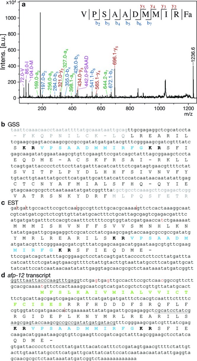
AF36 and afp-12. (a) MS/MS spectrum from ALA of AF36, m/z 1236.6. Peaks representing a (green), b (blue), y (red), and high-intensity internal fragment (purple) ions are labeled, and b and y ions are summarized in the sequence at the top of the spectrum. (b) Sequence from GSS database encoding AF36. (c) Sequence from EST library encoding AF36 (CB040272 (30)). (d) Cloned afp-12 transcript deduced by unique primers and 3′ RACE (HM125966). Gray indicates suspected intronic region. Blue indicates encoded peptide. Bold indicates basic cleavage sites. Green indicates signal peptide. Sequences used to design primers for PCR have a solid underline; the 5′ primer was SL1. Nucleotides with a dotted underline were used for the primer for 3′ RACE. In panels c and d red bases and vertical lines indicate insertions/deletions between the two sequences.