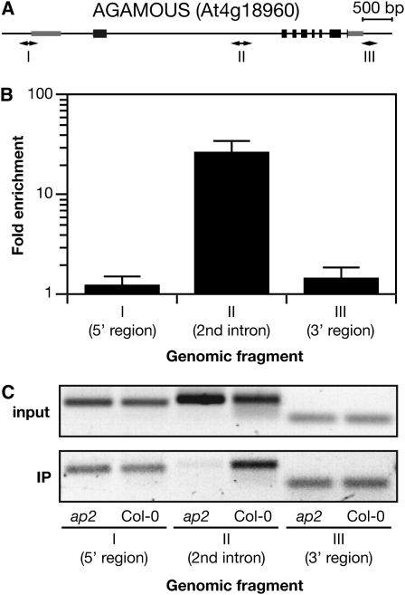
Figure 3.
AP2 Binds Directly to the Second Intron of AG.
(A) ChIP-qPCR. AG locus pictured with ChIP amplicons I, II, and III.
(B) qPCR analysis of triplicate biological replicate samples for binding enrichment of AP2 in the AG second intron. Replicates from independent experiments were measured to produce mean and 2xSEM for regions mapped in (A).
(C) Abundance of one biological replicate of the PCR products used to create (B). PCR product for input (top) and immunprecipitated (bottom) DNA is shown for Col-0 and ap2-12 for the three amplicons (I, II, and III) tiling the AG locus.

