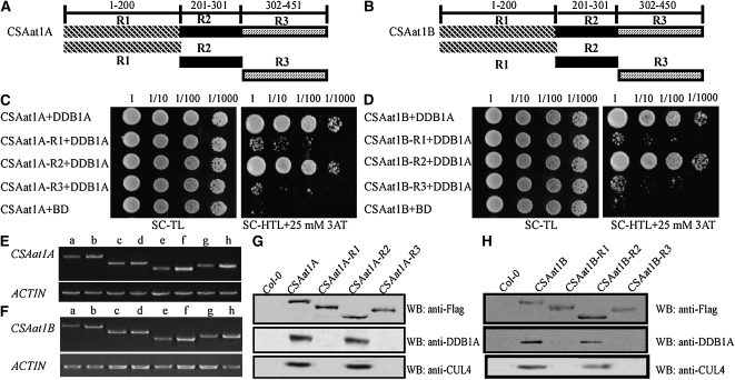
Figure 5.
The Second WD40 Domain in CSAat1A and B Interacts with DDB1A.
(A) and (B) Schematic representations of regions of the CSA1at1A and B proteins.
(C) and (D) Different regions of CSAat1A and B interact with DDB1A in yeast. Whole CSAat1A and B proteins as well as specific regions of the proteins (R1, R2, and R3) were cloned into pGADT7 and cotransformed with DDB1A into yeast. Growth of the transformed yeast was assayed on media minus Trp and Leu (left panels) or minus Trp, Leu, and His with 25 mM 3-amino-1,2,4-triazole (right panels). Columns in each panel represent serial decimal dilutions.
(E) and (F) Expression levels of CSAat1A and B and different fragments of the genes in their corresponding knockout mutants. a, c, e, and g, wild type; b, 35Spro-CSAat1A- or B-Flag; d, 35Spro-CSAat1A- or B-R1-Flag; f, 35Spro-CSAat1A- or B-R2-Flag; h, 35Spro-CSAat1A- or B-R3-Flag. RT-PCR products after 30 cycles with gene-specific primers and ACTIN as a loading control. Three replicate experiments were performed.
(G) and (H) Flag-tagged R1, R2, and R3 regions of CSAat1A and B interact with DDB1A and CUL4 in vivo. CSAat1A- and B-Flag proteins were immunoprecipitated with anti-Flag-agarose. The pull-down products were analyzed via immunoblots with anti-Flag, anti-DDB1A, or anti-CUL4 antibodies. WB, immunoblot.

