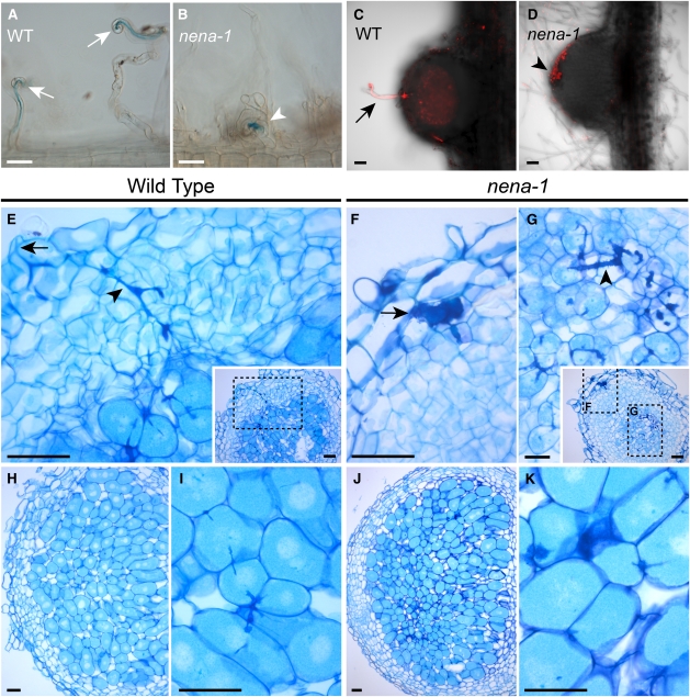
Figure 9.
Rhizobial Microcolonies at the Root Surface of nena-1 Lead to Nodule Formation and Intercellular Entry.
(A) and (B) Bright-field DIC images from roots hairs 7 DAI with lacZ-expressing M. loti and 18°C growth temperature. Wild-type (WT) plants show root hair curling (arrows) and ITs, whereas nena-1 mutants display abnormal root hair deformation and occasional colony formation by rhizobia (arrowhead). Images represent observations from more than eight plants per line. Bars = 50 μm.
(C) and (D) Confocal z-projections of longitudinal 80-μm tissue sections of a young infected wild-type (C) and an uninfected nena-1 (D) nodule. Images represent samples from 16 DAI/aerated (C) and 21 DAI/waterlogged + 5 μM AVG (D) treatments, corresponding to Figure 8D.
(C) DsRed expressing rhizobia (red) have colonized the nodule via an intracellular root hair IT (arrow).
(D) An uninfected nodule developed coinciding with accumulation of rhizobia at the root surface (arrowhead).
(E) to (K) Thin sections of nodule tissue stained with toluidine blue.
(E) Young wild-type nodule with intracellular IT (arrowhead) spanning from the infection site (arrow) into the cortex.
(F) and (G) Young nena-1 nodule with a subepidermal infection pocket (arrow) and cortical ITs (arrowhead).
Insets in (E) and (G) show respective sections at lower magnification; dashed boxes indicate magnified areas. Longitudinal sections of mature nodules from the wild type (H) or nena-1 (J) and corresponding magnifications ([I] and [K]) showing colonized host cells. Plants were grown under waterlogging conditions and sampled 3 WAI with M. loti R7A. Bars = 50 μm, except (G), where bar = 20 μm.

