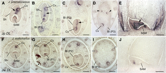
Figure 6.
Expression Pattern of DL in Leaf Primordia of J. prismatocarpus and J. wallichianus.
(A) to (E) In situ localization of Jp DL transcripts in J. prismatocarpus shoot apices.
(A) to (C) Transverse sections of shoot apices through the SAM (A), a P1 leaf blade (B), and a P2 leaf blade (C), showing strong Jp DL expression in the central domain of leaf primordia (arrows).
(D) Transverse section through a P3 leaf blade, showing Jp DL expression in the secondary central domain (arrowheads).
(E) Longitudinal section through the SAM showing strong Jp DL expression (arrow).
(F) to (J) In situ localization of Jw DL transcripts in J. wallichianus.
(F) to (I) Transverse sections of shoot apices through the SAM (F), a P1 leaf blade (G), a P2 leaf blade (H), and a P3 leaf blade (I), showing no Jw DL expression in the mesophyll tissues and weak expression around the central vascular bundle. White arrowheads indicate loss of Jw DL expression in the secondary central domain.
(J) Longitudinal section through the SAM showing weak Jw DL expression.
Bl, leaf blade; Sh, leaf sheath; Cv, central large vascular bundle. Plastochron numbers of leaf blades are indicated (P1, P2, and P3). Note that the central large vascular bundle differentiates in a slightly off-center position in Juncus leaves. Bars = 200 μm.

