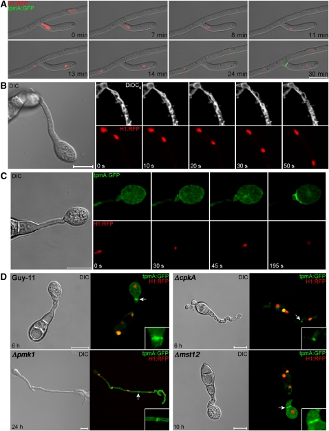
Figure 1.
Spatial Uncoupling of Mitosis and Cytokinesis during Infection-Related Development in the Rice Blast Fungus M. oryzae.
(A) Laser confocal micrographs of a time series to show actomyosin ring formation in M. oryzae expressing tpmA:GFP and histone H1:RFP during vegetative hyphal growth. The site of septation in a hyphal branch occurs at the medial position of the preceding nuclear division.
(B) Time series of micrographs showing mitosis occurring during appressorium development by M. oryzae. Conidial suspensions of the M. oryzae H1:RFP strain were prepared and the lipophilic stain DiOC6 used to stain the nuclear envelope. A differential interference contrast (DIC) image of the whole germ tube and developing appressorium is shown in the left panel.
(C) Time series to show actomyosin contractile ring formation during differentiation of the appressorium in M. oryzae tpmA:GFP-Histone H1:RFP strain. Left panel shows DIC image of the nascent appressorium. Right panels show TpmA:GFP and H1:RFP signals, respectively.
(D) Micrographs of M. oryzae strain Guy-11, ΔcpkA, Δpmk1, and Δmst12 mutants expressing H1:RFP, and tpmA:GFP gene fusions incubated on cover slips to allow appressorium development. Septation was spatially separated from the site of nuclear division only in Guy-11 and the Δmst12 strains, which are competent in appressorium formation. All images were recorded using a Zeiss LSM510 Meta laser confocal laser scanning microscope system.
Bars = 10 μm.

