Fig. 7.
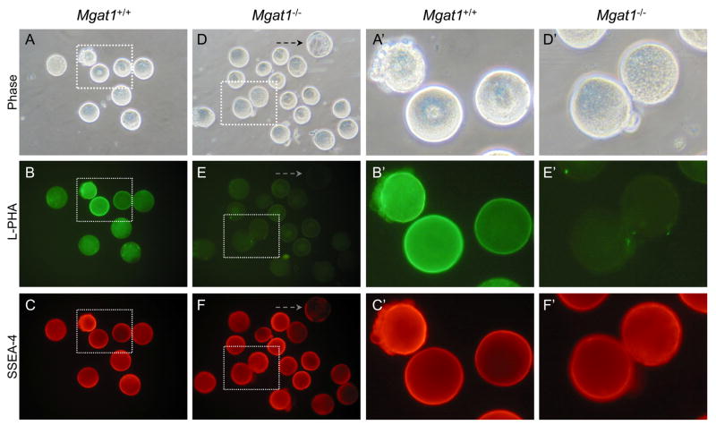
SSEA-4 detection on ovulated eggs from Mgat1 mutant females. Mgat1FF:ZP3Cre females were superovulated and zona-free eggs prepared. A late wild type blastocyst (dashed arrow) was included with the mutant eggs as a negative control for SSEA-4. A. Phase contrast of control zona-free eggs obtained from superovulated Mgat1F/F females. B. L-PHA binding to control eggs in A. C. SSEA-4 MAb binding to control eggs in A. D. Phase contrast of zona-free eggs obtained from Mgat1F/F:ZP3Cre females. E. L-PHA binding to the Mgat1 mutant eggs in D. F. SSEA-4 MAb binding to the mutant eggs in D. L-PHA negative eggs fluoresce strongly. A′-F′. The area marked by each dotted square in A-F is magnified. Images are at the same magnification and the same exposure. Images are representative of 2 experiments with 10 eggs and 4 compacted 8-cell embryos, half of the total generated by 7 control females, and 16 eggs and 6, 4–8 cell embryos, half of the total generated by 5 Mgat1 mutant females.

