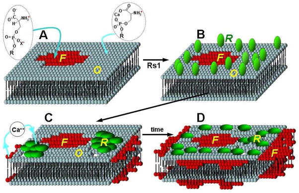
Fig. 8.
Schematic illustration of the hypothesized interaction of Rs1 with PS in the presence of Ca2+. A. Ca2+ controlled PS phases before Rs1 introduction; The PS bilayer exists in two phases, a fluid phase devoid of calcium (“F”) and a calcium-rich ordered phase (“O”). B. Rs1 adsorbs randomly onto the ordered lipid phase. Electrostatic interactions lead the protein to preferentially adsorb to the ordered lipid domain. Adsorption is followed by protein insertion toward the hydrophobic lipid core causing lipid disruption and apparent expansion of the fluid phase. C. At this point the protein assembles into labile patches. D. These patches tend to diffuse toward a more homogeneous distribution over time. Anchoring of the protein to the lipid enhances the membrane robustness significantly. This is consistent with the role of Rs1 as an adhesion molecule that helps maintain the architectural integrity of the retina. “F” fluid phase (Ca2+-poor); “O” ordered Phase (Ca2+-rich); “R” Rs1 protein;

