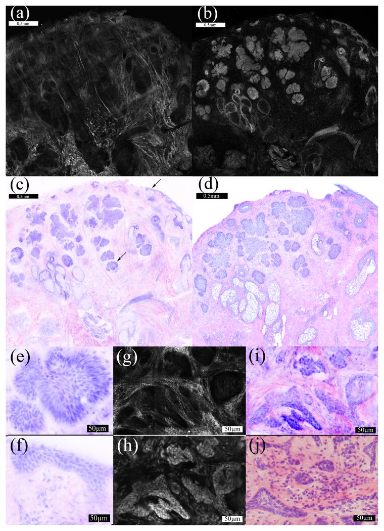
Figure 1.
(A–D) 2X magnification. The reflectance mosaic (a) is colored pink and the fluorescence mosaic (b) is colored purple in the DSCP image (c). The correlating H&E histopathology (d) is also shown at 2X magnification. The arrows (c) indicate a micronodular focus in the deeper dermis and the superficial epidermis. (e–j) 30X magnification. A micronodular tumor focus in the dermis (e) and normal superficial epidermis (f) are magnified from figure 1c. In a separate sample containing infiltrative collagen is bright in the deeper dermis in reflectance mode (g). The correlating fluorescence image (h) shows bright nuclei. The DSCP image (i) is shown with the correlating histopathology (j).

