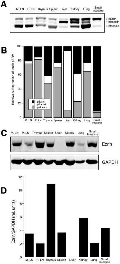
Figure 1. Expression of ezrin and moesin in lymphoid organs.
Mouse organs were lysed in SDS sample buffer and analyzed by western blot with antibodies specific for pERM (A), and ezrin or GAPDH (C). B. Relative abundance of each pERM protein within a tissue was calculated using an Odyssey Imager and normalized to the total pERM expression in that tissue. D. Expression of ezrin among tissues was calculated after normalization to GAPDH.

