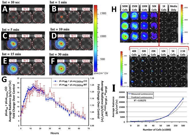
Figure 2. in vitro bioluminescent imaging of lux cassette containing cells.
pLuxCDEfrp:CO/pLuxAB containing (CO), pLuxCDEfrp:WT/pLuxAB containing (WT), and untransfected negative control (NEG) HEK293 cells were plated in 24-well tissue culture plates and integrated for (A) 10 sec, (B) 1 min, (C) 5 min, (D) 10 min, (E) 15 min, and (F) 30 min. Bioluminescence from cells co-transfected with pLuxCDEfrp:CO/pLuxAB was distinguishable from background in the presence of untransfected cells after 10 sec and showed no increase in background detection even after a 30 min integration time. Long term in vitro expression (G) demonstrates the temporal longevity of the signal without exogenous amendment. The minimum detectable number of bioluminescent cells was determined (H) by plating a range of cell concentrations in equal volumes of media in triplicate (downward columns) in an opaque 24-well tissue culture plate. The minimum number of cells that could be consistently detected was approximately 20,000. Average radiance was shown to correlate with plated cell numbers (I), yielding an R2 value of 0.95275.

