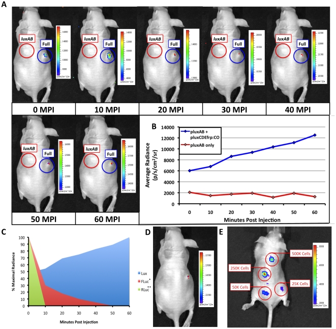
Figure 3. in vivo bioluminescent imaging using HEK293 cell expression of mammalian-adapted lux.
(A) HEK293 cells containing the mammalian adapted pLuxCDEfrp:CO/pLuxAB cassette (Full) were subcutaneously injected into nude mice and imaged. Detection occurred nearly immediately (<10 sec) post-injection and remained visible up to the 60 min time point of the imaging assay. HEK293 cells containing only the pLuxAB plasmid (luxAB) were subcutaneously injected into the same mouse as a negative control. Note that the automatic scaling of signal intensity differs among images, therefore creating the false appearance that image intensity is decreasing after the 10 min post-injection time point when in fact it continually increases as shown in panel (B). (C) Comparison of mammalian-adapted lux-based bioluminescence from HEK293 cells versus published data on the expression of FLuc (*[25]) and RLuc (**[8]) tagged cells over the 60 min course of the assay. (D) Upon termination of the assay 60 min post injection, the bioluminescent signal from HEK293 cells expressing the full complement of lux genes was detectable using an integration time as low as 30 sec. (E) Subcutaneous injection of HEK293 cells containing pLuxCDEfrp:CO/pLuxAB at concentrations ranging from 500,000 to 25,000 cells in 100 µl volumes of PBS demonstrated a tested lower limit of detection of 25,000 cells using a 10 min integration time. MPI, minutes post injection.

