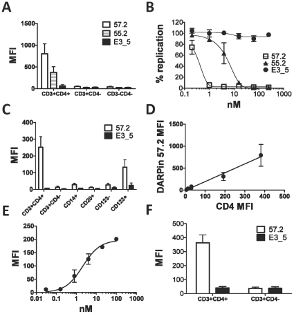
Figure 1. CD4-specific DARPin 57.2 binds to macaque CD4+ cells.
A. PBMCs were incubated with 200nM of DARPins 57.2, 55.2 or E3_5 for 30 minutes before being labeled with antibodies for CD3, CD4 and His-tag. Mean fluorescent intensities (MFIs) of the anti-His staining on the indicated subsets identified within a total leukocyte gate from 4 independent experiments are shown (mean±SEM). B. PBMCs were pretreated with the indicated concentration of DARPins 57.2, 55.2 or E3_5 and then exposed to SIVmac239. 7 days later the amount of p27 in the culture supernatant was quantified by ELISA. The values shown are the means (±SEM) from 3 independent experiments and are normalized to the amount of p27 produced in the absence of inhibitors (i.e. 100% replication). C. PBMCs were incubated with 200nM of DARPins 57.2 or E3_5 for 30 minutes before being labeled with antibodies for CD3, CD4, CD14, CD20, CD123, HLA-DR and His-tag, as indicated. MFIs (mean±SEM) of the anti-His staining on the indicated subsets identified within a total leukocyte gate from 5 independent experiments are shown. The DC-containing fractions were identified within the Lineage−HLA-DR+CD123− (CD123−, myeloid DC-containing fraction) or the Lineage−HLA-DR+CD123+ (CD123+, plasmacytoid DCs). D. The different cell types defined in panel C were stained with anti-CD4 (in the absence of DARPin 57.2) or anti-His (in the presence of DARPin 57.2) in parallel and the CD4 MFIs determined (Table 2). Each point on the graph represents a distinct cell type. Linear regression and correlation were performed using the GraphPad Prism software. An average of MFIs of the anti-His (DARPin) and anti-CD4 staining for each indicated cell type from 3 independent experiments is shown. E. PBMCs were incubated with various concentrations of DARPin 57.2, labeled with the anti-penta His antibody and analyzed by flow cytometry. MFIs (mean±SEM) from 2 independent experiments are shown. Curve fitting was done using the GraphPad Prism software. F. Lymph node cells were incubated with 200nM of the DARPin 57.2 or E3_5 and stained for His and CD4. MFIs (mean±SEM) of the anti-His staining of the gated CD4+ cell population from 5 independent experiments are shown.

