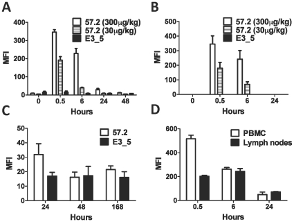
Figure 4. Injection of macaques with DARPin 57.2 results in transient binding to CD4+ cells.
A. Blood samples were taken from macaques at indicated timepoints after an injection with DARPin. PBMCs were isolated, labeled with antibodies to CD4 and to penta-His and analyzed by flow cytometry. The MFI (mean±SEM) of His staining on CD4+ cells within a total leukocyte gate is shown for animals injected with 300µg/kg DARPin 57.2 (n = 6), 30µg/kg DARPin 57.2 (n = 3) or DARPin E3_5 (n = 5). Data from both doses of DARPin E3_5 was combined for simplicity. B. Whole blood samples taken after DARPin injection were immediately labeled with the anti-His antibody and analyzed by flow cytometry. The MFIs (mean±SEM) of His+ cells are shown for animals injected with 300µg/kg DARPin 57.2 (n = 6), 30µg/kg DARPin 57.2 (n = 3) or DARPin E3_5 (n = 5). Lymph nodes (C and D) and blood (D) were taken from macaques at indicated timepoints after an injection with DARPin. Isolated cells were labeled with antibodies to penta-His and CD4 and analyzed by flow cytometry. MFIs (mean±SEM) of gated CD4+ cells are shown for animals injected with 300µg/kg DARPin 57.2 (C, n = 7; D n = 4) or DARPin E3_5 (C, n = 6).

