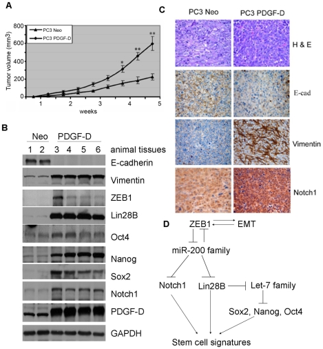
Figure 8. The cells with EMT phenotype promoted tumor growth.
(A) Tumor growth curve showing that over-expression of PDGF-D could promote tumor growth in SCID mice much faster than PC3 Neo cells. (B) Western blot analysis of tumor lysates showing the expression of EMT and stem cell makers. (C) H&E evaluation of the tumors from both the groups showed high grade carcinoma associated with tumor apoptosis and necrosis. The results from the immunostaining showed much intense staining for vimentin and Notch1 and less intense staining for E-cadherin. (D) Regulatory model showing mechanistic link between EMT and stem cells: EMT induced by different factors characterized by increased expression of ZEB1, which causes loss of miR-200 family. Loss of miR-200b and miR-200c lead to increased ZEB1, Notch1 and Lin28B expression, resulting in decreased expression of let-7. Downregulation of let-7 leads to the up-regulation of Sox2, Nanog and Oct4, which together with Notch1 and Lin28B contribute to stem cell signatures. (*, p <0.05, **, p<0.01 compared to PC3 Neo, E-cad: E-cadherin).

