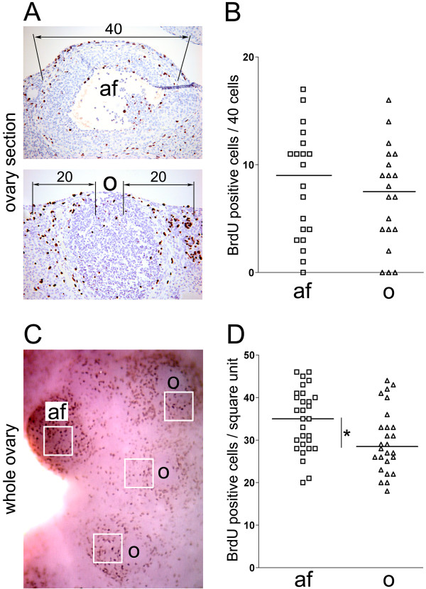
Figure 2.
Quantification of BrdU incorporation during antral follicle formation and ovulatory wound repair in ovary sections (A, B) and whole ovaries (C, D). (A) In ovary sections, the number of BrdU positive cells associated with the antral follicles (af) was determined by scoring forty cells covering the surface of each antral follicle. The number of BrdU positive cells associated with corpora lutea containing healing ovulatory wounds (o) was determined by scoring twenty cells covering each side of the ovulatory wound. (B) Dot plot of the number of BrdU positive cells associated with antral follicles and ovulatory wounds. Each rectangle represents one antral follicle (N = 20) and each triangle represents one corpus luteum with an ovulatory wound (N = 20). The horizontal bars represent the mean values. P = 0.5026. (C) In the whole ovary, the number of BrdU positive cells associated with antral follicles (af) and ovulatory wounds (o) was determined by counting positive cells per square unit. (D) Dot plot of the number of BrdU positive cells associated with antral follicles (af) and ovulatory wounds (o). Each rectangle represents one antral follicle (N = 29) and each triangle represents one corpus luteum with an ovulatory wound (N = 26). The horizontal bars represent the mean values. *P = 0.0175.

