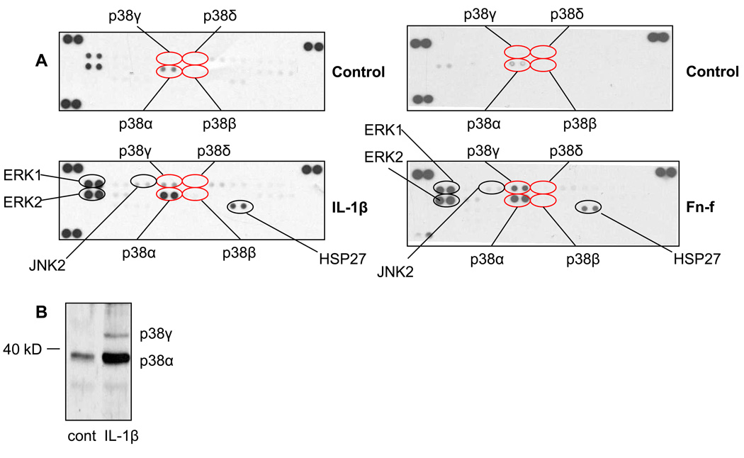
Fig. 1. p38α and p38γ are phosphorylated in chondrocytes following catabolic stimulation.
(A) Chondrocytes were stimulated for 30 minutes with either 10ng/mL IL-1β or 500nM Fn-f or with control media. Lysates were analyzed on a MAPK antibody array. Results are representative of three independent experiments. (B) Chondrocytes were stimulated with 10ng/mL IL-1β. Lysates were immunoblotted with pan phosphospecific p38 antibody that recognizes all isoforms. Anticipated locations of p38γ (above 40kD) and p38α (below 40kD) are marked. Results are representative of three independent experiments.

