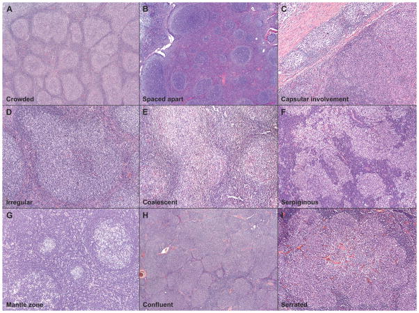
Figure 2. Spectrum of Follicle Morphology.
Neoplastic follicles in follicular lymphoma showed either crowded (A) or spaced-apart (B) follicles, and many also showed involvement of the capsule (C). In addition, the follicles also showed wide variation in the shape and size: typical examples of irregular (D), coalesced (E), serpiginous (F), mantle zone differentiation (G), confluent (H), and serrated (I) follicles are illustrated.

