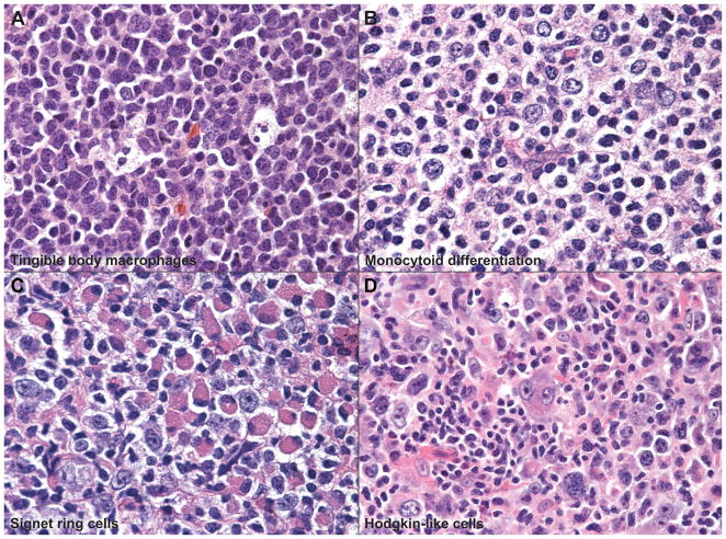
Figure 3. Spectrum of Cytologic Features.
The cellular composition and cytologic features of the neoplastic follicles showed variation and include: (A) follicles with tingible body macrophages, (B) monocytoid differentiation, (C) signet-ring like features with eccentric nuclei and abundant cytoplasm or cytoplasmic inclusions, and (D) a rare example of FL with Hodgkin/Reed-Sternberg-like cells within follicles.

