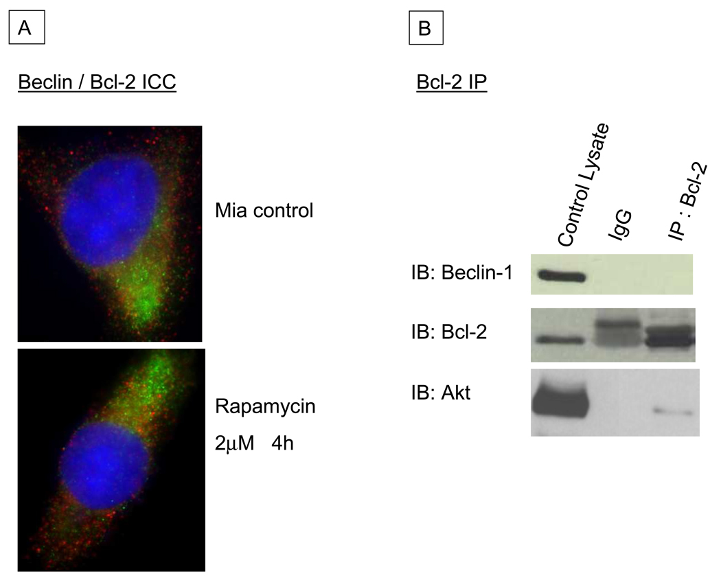
Figure 5.
A: Immunocytochemistry of normal MIA-PaCa-2 cells (top) and those treated with Rapamycin to induce autophagy (bottom), labeling Beclin 1 (green) and Bcl-2 (red). Minimal to no co-locolization of the signals is observed. Nuclei are stained blue with 4,6-diamidino-2-phenylindole dihydrochloride (DAPI). B: Immunoblots of normal MIA-PaCa-2 cell lysates following immunoprecipitation (IP) with a monoclonal Bcl-2 antibody. The left-hand lane contains lysate prior to IP, the middle lane is a control for non-specific binding showing IP with a non-specific IgG, and the right-hand lane contains the Bcl-2 IP fraction. No Beclin 1 is seen in the Bcl-2 immunoprecipitated fraction, but Akt is detected as a positive control for the IP assay.

