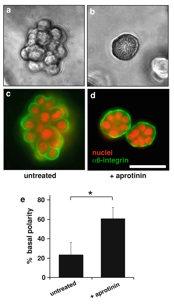
Fig. 1.
Effect of serine protease inhibitor aprotinin on growth morphology of T4-2 cells in 3D culture. Cells were cultured in Matrigel for 8 days either in the absence (panels a, c) or in the presence (panels b, d) of 1 mg/ml aprotinin. Aprotinin suppressed disorganized growth and led to formation of acini with basal polarity, as demonstrated by phase contrast microscopy (a, b), and by immunofluorescence staining for α6 integrin (c, d). Scale bar, 50 µm. e Quantitative analysis of polarity by percentage of colonies with polarized distribution of basal α6 integrin confirmed that a significantly greater proportion of colonies showed basal polarity in the aprotinin-treated culture. Data are expressed as mean ± SD. * P < 0.01 (unpaired t test)

