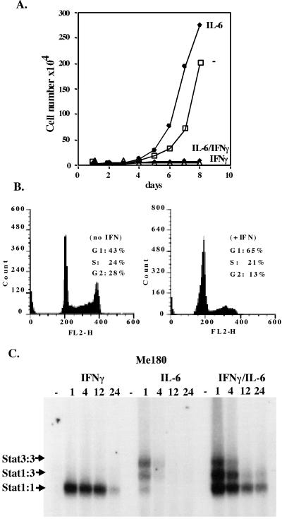
Figure 2.
Me180 cells are growth inhibited by low concentrations of IFNγ. (A) 1 × 10⁴ Me180 cells were plated onto 3-cm dishes. Once the cells had adhered (≈12 h later), IFNγ (0.5 ng/ml), IL-6 (200 units/ml), both, or neither were added to the adherent cells grown in complete media. On a daily basis, the cells were counted by trypan blue exclusion (the mean of triplicate samples). (B) 1 × 10⁶ cells were plated onto 6-cm dishes and treated with or without IFNγ (0.5 ng/ml) for 24 h. The cells were harvested, stained with propidium iodide (PI), and analyzed by FACS. The number of cells in G1, S, and G2 was determined. (C) Me180 cells were grown and treated with either IFNγ (0.5 ng/ml), IL-6 (200 units/ml), or both for 1, 4, 12, or 24 h. Cells were harvested and nuclear extracts were isolated from the cell samples. Equal amounts of protein were incubated with radiolabeled double stranded m67(binding site for Stat1 and or Stat3) and resolved on a nondenaturing acrylamide gel.

