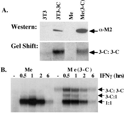
Figure 3.
Me180 cells expressing Stat3-C. (A) Stable clones of Me180 cells were isolated expressing either a control plasmid or a Stat3-C expressing plasmid. The clones were maintained in G418 containing media. Nuclear extracts were isolated from these clones and one example is shown here. Ten micrograms of protein from each cell line (3T3, 3T3–3C, Me, Me3-C) was resolved on an SDS polyacrylamide gel, transferred to nitrocellulose, and probed with anti-FLAG antisera. Two micrograms of nuclear extract from the same cell lines (see above) were incubated with radiolabeled m67 and resolved on a nondenaturing EMSA gel. (B) Parental Me180 cells (Me) and Me180 expressing Stat3-C cells (Me 3-C) were either left untreated (serum containing media) or treated with IFNγ (0.5 ng/ml) for 0.5, 1, 2, or 6 h. Nuclear extracts were isolated from these, incubated with radiolabeled m67, and resolved on an EMSA gel.

