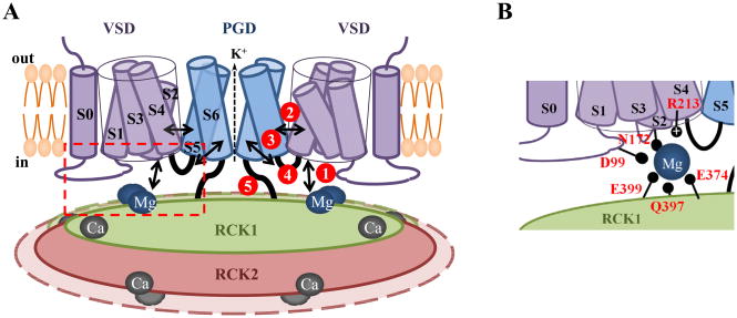
Figure 2.
Interactions between structural domains in BK channels. A) Cartoon of the BK channel. Mg2+ and Ca2+ show metal binding sites. Black arrows and heavy set lines are used to indicate the following interactions: 1) between VSD and RCK1 in Mg2+ dependent activation; 2) between VSD and PGD through S4 and S5; 3) between S4-S5 linker and S6; 4) the tug of the S4-S5 linker and 5) between PGD and cytosolic domain through the peptide C-linker. The gating ring formed by RCK1 and RCK2 is shown to undergo expansion during channel gating similar to the MthK channel from Methanobacterium thermoautotrophicum. The structure in dashed box is shown in more details in B. B) Cartoon showing residues involved in Mg2+ dependent activation.

