Figure 4.
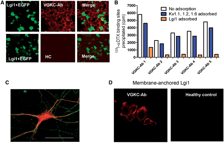
Lgi1-antibodies are present in many VGKC-antibody positive patients. (A) HEK293 cells were co-transfected with cDNAs for Lgi1 and EGFP (green) and incubated with patient sera (1:20). A representative image of binding of a limbic encephalitis VGKC-antibody positive serum to the cells, detected with anti-human IgG (red). Note that the red stain is found not only on the Lgi1/EGFP transfected cells, but also on the surface of non-transfected cells. Similar results were found in 46 of the 96 patient sera, but were not observed with healthy control sera (HC). (B) Immunoadsorption of the VGKC-antibody positive sera against Lgi1-transfected cells, but not against Kv1-transfected cells, substantially reduced immunoprecipitation of 125I-α-DTX–VGKC complexes from rabbit brain extracts. (C) Lgi1-positive sera binding to a live hippocampal neuron detected with fluorescein-anti-human IgG (green); the neurons were subsequently fixed, permeabilized and stained for MAP2 (red). The bar represents 25 µm. (D) To confirm that Lgi1 was the target for the antibody binding shown in A, cDNA for Lgi1 was fused to cDNA for the transmembrane domain and cytoplasmic tail of Caspr2. This construct expressed well in HEK293 cells and provided a direct assay for Lgi1 antibodies. Nine additional sera (one illustrated here) bound to these cells; but there was no binding of healthy control sera or Caspr2-antibody positive sera.

