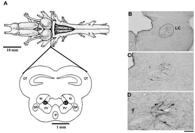
Figure 5.
A. Dorsal view of the anuran brain, indicating the level of midbrain section illustrated below. Adapted from Gargaglioni and Branco (2009). B, C and D. Photomicrographs of the isthmus region showing the catecholaminergic cell bodies identified by tyrosine hydroxylase immunohistochemical staining of the brains. Detailed (higher magnification) morphology of these neurons is shown in B and C. Scale bars: 200 μm in A, 100 μm in B, and 50 μm in C. Adapted from Noronha-de-Souza et al. (2004). Abbreviations: Aq, Aqueduct of Sylvius; LC, Locus coeruleus; NI, nucleus isthmi; Otec, optic tectum. LC, locus coeruleus; IV, fourth ventricle.

