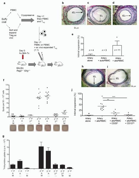
Figure 2. TA mediated by allogeneic human PBMC in human arterial interposition grafts is attenuated with human Treg cells.
(a) A schematic representation of the in vivo protocol. (b–d) EvG staining of a human artery procured from mice transplanted with an artery alone and not reconstituted with PBMC (b); or reconstituted with either 10 × 106 allogeneic human PBMC (c) or 10 × 106 autologous PBMC taken from the arterial donor (d). (e) Percentage of intimal expansion in human arteries from all experiments. (f) Results of ELISPOT for human IFN-γ and (g) MLR performed with cells from both the primary (1°) and secondary (2°) cultures (f and g, 2 independent experiments with ≥ 4 replicates; P denotes responding PBMC, A – irradiated allogeneic PBMC, 3rd – irradiated 3rd party PBMC, M – irradiated mouse splenocytes). (h,i) EvG staining of human artery procured from mice inoculated with PBMC + CD25hi Treg cells (h) or PBMC + CD127lo Treg cells (i) at a 1:1 ratio (10 × 106 cells). Histological quantification of intimal expansion in mice treated with Treg cell therapy (j). Error bars are represented as means ± s.d. * P < 0.02 in the artery + allo-PBMC group when compared to controls, ** P = 0.0013, ***P = 0.0007. IEL denotes internal elastic lamina.

