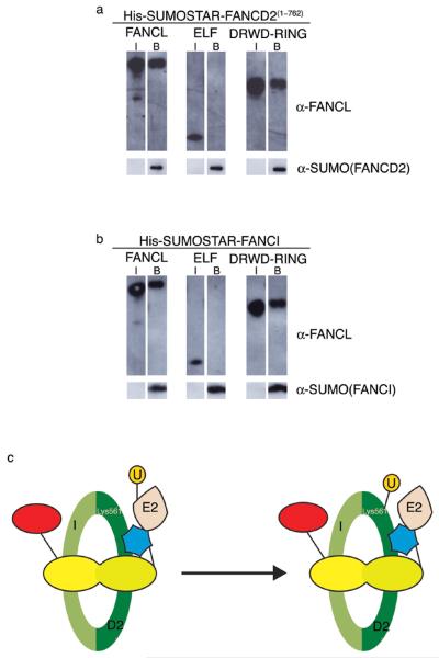
Figure 4. The DRWD domain recruits substrate.
a. Pulldown binding assays using His-SUMOSTAR-FANCD2(1–762) as bait. Input samples (I) of FANCL species and samples bound by His-SUMOSTAR-FANCD2(1–762) (B) are indicated. Samples are analysed by Western blotting using anti-FANCL or anti-SUMO antibodies. b. Pulldown binding assays using His-SUMOSTAR-FANCI as bait. Input samples (I) of FANCL species and samples bound by His-SUMOSTAR-FANCI (B) are indicated. Samples are analysed by Western blotting using anti-FANCL and anti-SUMO antibodies. c. A schematic representation of FANCD2 monoubiquitination. The I/D2 complex is represented in green, with Lys561 indicated. The ELF, DRWD and RING domains are red, yellow and blue, respectively. The E2 and ubiquitin (U) are indicated.

